Biopsy
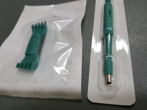
Shave biopsy (left) and punch biopsy (right)
If your doctor does a skin check and finds any spots that look concerning, they will likely recommend a biopsy.
A biopsy usually involves cleaning the area and injecting some local anaesthetic to numb the skin.
They will then either take some or all of the concerning spot and this will be sent to the lab where it will be closely looked at by specialist pathologists who will send a report back to your doctor regarding the spot.
By doing a biopsy your doctor is able to determine what a particular spot is and then work out the most appropriate way to treat it.
There are a few different methods for taking a biopsy and these mostly depend on what is being biopsied, it’s location and your doctors skills + expertise.
If you have a dark spot and your doctor is concerned about melanoma they will generally aim to take the whole spot.
- This can either be done by cutting out the spot and stitching the skin back together or can sometimes be done by using a flexible razor blade that “shaves” the spot out of your skin.
Excision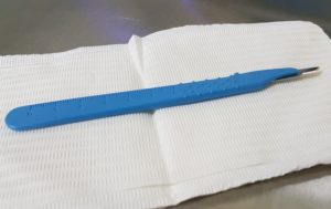
Most skin cancers can be treated by excision which usually involves the following:
- The area is cleaned well and local anaesthetic is injected to numb the area.
- The skin cancer is then cut out, usually with a scalpel – this is often done in an ellipse shape to allow the skin to come back together with the least tension.
- The spot that has been cut out is then sent to the lab where it will be closely analysed to ensure that the entire spot has been cut out – if it isn’t all cut out then sometimes this will require another excision at the affected area.
- Sometimes your doctor may use technique to minimise bleeding at the area.
- The area is then closed by stitching – most often these stitches aren’t absorbable and they will usually need to be removed in 5-14 days depending on the body area.